
Our Research
A central challenge in neuroscience is identifying and manipulating specific cell types in the brain to study their function. The Chen Lab addresses this by creating precise genetic tools that target individual neuron types across all developmental stages in Drosophila.
Decoding the Molecular Logic of Sensory Circuit Assembly
During neuronal development, sensory neurons acquire the correct cell fates and connect to the proper partners. The formation of these functional circuits is essential for animals to detect environmental input and drive behavior. Yet, dissecting the molecular logic underlying sensory circuit formation in higher-order organisms remains challenging, in part due to the complexity and diversity of their nervous system.
We address this challenge using Drosophila and the precise genetic tools we build in the lab (see Our Unique Approach) to identify and manipulate specific cell types at the level of individual circuits.
Major Scientific Questions We’re Investigating:
-

Wiring the Visual Brain: Finding the Right Target
-
Keeping the Picture Straight: Precision in Visual Mapping
-

How Chance Becomes Order: Wiring a Randomized Circuit
Key Research Projects
Single neuronal labeling of TmY14 cell in the fly visual system.
Molecular regulation of visual circuit assembly
Uncovering the Molecular Controls of Neuropil Targeting in Developing Sensory Circuits
How do developing neurons know where to extend their branches in the brain? Understanding how immature neurons develop specific structures that allow them to connect with precise brain regions is a key question in our lab. We are using the TmY14 neuron in the Drosophila visual system to tackle this challenge. TmY14 neurons extend branches to multiple regions — the medulla, lobula, lobula plate, and the central brain. We have identified candidates for three transcription factors that might regulate these projections during development. Our ongoing work focuses on unraveling the molecular network involving these transcription factors and identifying the downstream signals that direct neuronal targeting and circuit formation.
Development and maintenance of retinotopic map
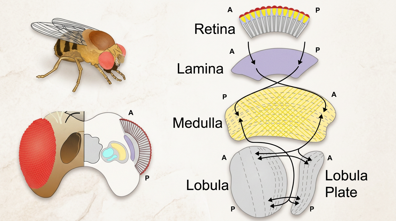
The Drosophila visual system preserves retinotopic order across sequential optic lobe neuropils. Photoreceptors in the retina project to the lamina, medulla, lobula, and lobula plate in a point-to-point map that maintains the anterior–posterior (A–P) axis, with a single axial inversion between the lamina and medulla.
Maintaining the Retinotopic Map: Decoding Precision in Visual Circuit Wiring
Imagine a perfectly aligned grid, where each point in the eye corresponds exactly to a point in the brain. This precise organization, called retinotopy, is essential for maintaining spatial accuracy in visual processing. But how are these connections established during development?
Our lab investigates this question using the Y3 neuron in the Drosophila visual system. During early pupation, Y3 neurons send projections to the lobula and lobula plate but only extend their branches to the medulla later in development. This delayed targeting suggests that more than just birth order governs connection specificity.
We aim to identify candidate molecular regulators using Y3 developmental transcriptomes and to test how these molecules influence Y3 neurite targeting in the medulla. Additionally, we will assess whether other cell types act as “guiding wires” by selectively ablating them and observing the impact on Y3 neuron targeting. Our goal is to reveal how a precisely organized grid of cells is constructed during development, ensuring that visual information is accurately mapped from the retina to the brain.
Development of a Stochastic Circuit
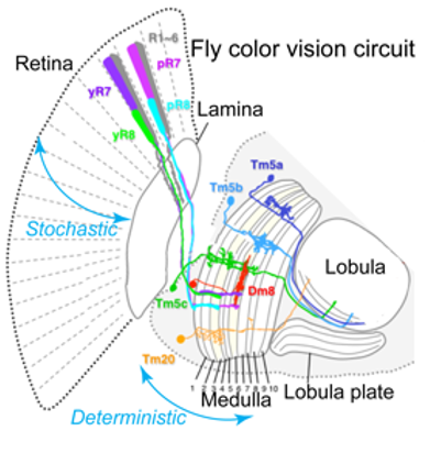
Schematic of the color vision
circuit with
axonal projections of the
four stochastically
specified photoreceptors
and their
downstream targets in the medulla. Modified
from Song BM, Lee CH. Front Neural Circuits. 2018. PMID: 29527156.
Identifying Molecular Controls of Stochastic Color Vision Circuit Assembly
Previous work in my postdoc lab (Claude Desplan’s lab) demonstrated that stochastic color photoreceptors (yR7) connect to deterministic partners in the medulla (yDm8) through two immunoglobulin superfamily (IgSF) proteins (Dpr11-DIPγ). During my postdoc, I identified another IgSF pair from the Beat and Side family, guiding the pale counterparts (pR7 to pDm8). This y/p information needs to be propagated throughout the circuit.
The main outputs of Dm8 are projection neurons that connect to the lobula in the optic lobe. It is unclear how the y/p organization of the eye (R7/R8) and medulla (y/pDm8) is preserved in the lobula neuropil or the central brain. We will use published EM connectome data to identify the targets of y/p-specific projection neurons and examine the y/p columnar organization in the lobula. We will also prevent the generation of y or pR7s (using Ss gain- or loss-of-function) and generate scRNAseq libraries to determine which neural types depend on the presence of y/p neurons. Our work aims to uncover how stochastic decisions propagate to downstream partners at the system level.
resource building for the neuroscience community
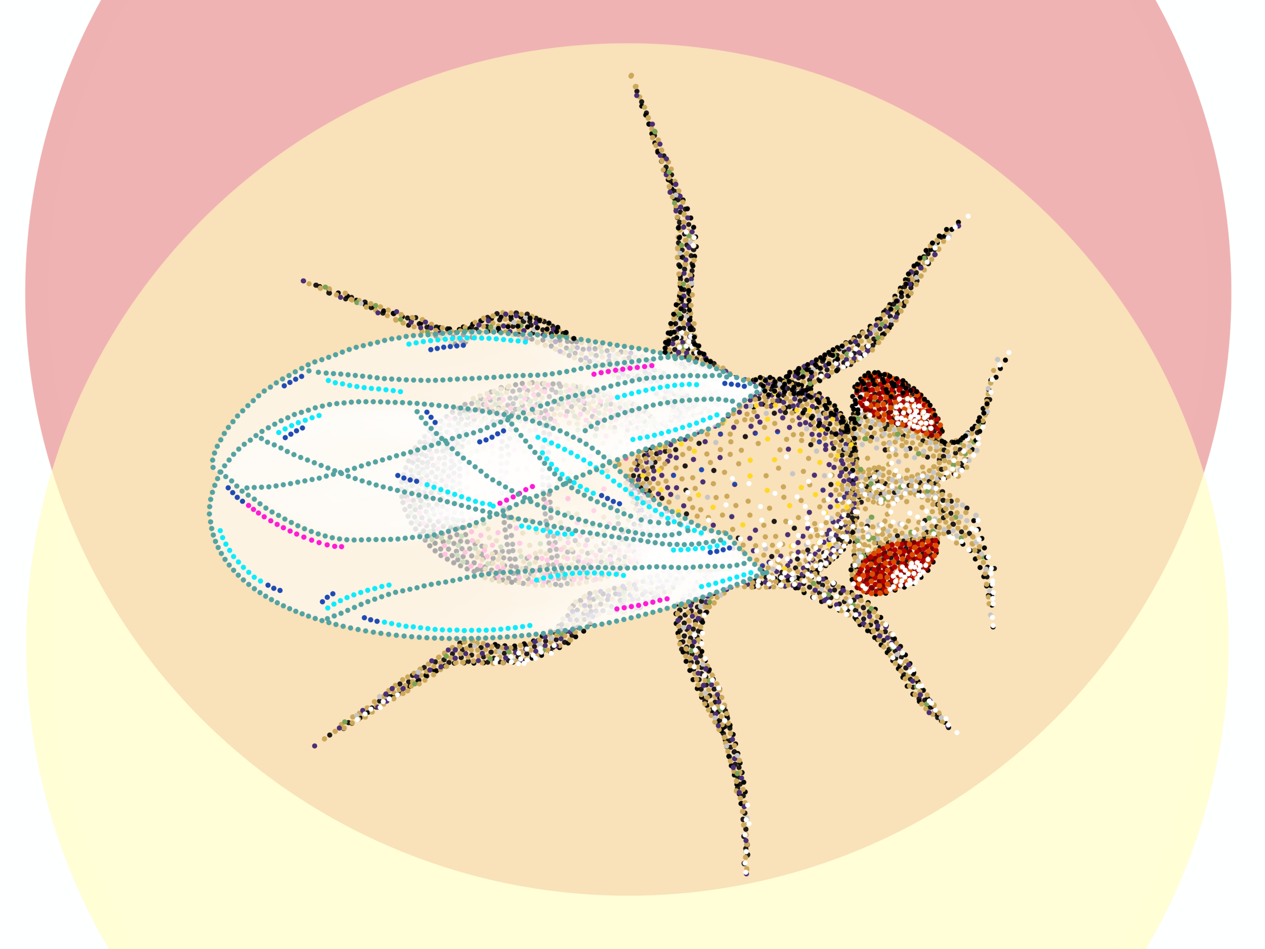
Single-cell transcriptomics guides intersectional genetic targeting of developing visual neurons in Drosophila.
Each dot represents a single-cell transcriptomic cluster from the developing fly visual system. The overlapping ovals illustrate the logic of the split-GAL4 system, in which co-expression of two hemi-drivers (DBD and AD) restricts transgene expression to a precise genetic intersection. By matching cluster-specific marker genes to split-GAL4 driver combinations, we generate cell-type-specific tools to label, manipulate, and characterize individual neuron populations as they emerge during visual system development. Image credit: Veronica Hughes, STEAM visuals.
Expanding the Genetic Toolkit to Decode Brain Wiring
How do we identify and manipulate specific cell types in the brain to study their function? Our lab addresses this challenge by creating precise genetic tools that allow us to target specific neuron types across all developmental stages in Drosophila.
Our approach leverages single-cell RNA sequencing (scRNAseq) data to develop gene-specific split-GAL4 lines. Unlike existing enhancer-based genetic tools that often lack specificity and may not work consistently throughout development, our method focuses on regulatory elements identified through scRNAseq. This results in highly predictive, reliable tools for targeted genetic manipulation.
During his postdoc, Dr. David Chen led a team of undergraduates to streamline the protocol for generating split-GAL4 lines (Li & Li et al., 2023 Star Protocol). Using this protocol, approximately 3,000 fly lines from the Drosophila Stock Center can be converted into split-GAL4 lines, facilitating precise targeting of cell types across various tissues. Our lab actively collaborates with researchers at Columbia, UPenn, and the University of Toulouse to expand the toolkit and apply it to diverse developmental and functional studies in Drosophila neurobiology.
Join the lab!
Are you someone who loves to ask questions, solve puzzles, and learn new techniques?
Whether you’re interested in genetics, neurobiology, or computational analysis, our lab offers a welcoming and supportive environment where you can develop your skills while contributing to groundbreaking research. We’re actively recruiting undergraduates, graduate students, and postdocs who are passionate about understanding how the brain builds itself, one neuron at a time.
Reach out to discuss research opportunities and find out how you can be part of the team!